L1R Antibody

Code CSB-PA324949ZA01VAA
Size US$299
Image
  • Western Blot
    Positive WB detected in Recombinant protein
    All lanes: L1R antibody at 1:2000
    Secondary
    Goat polyclonal to rabbit IgG at 1/50000 dilution
    Predicted band size: 26.8 kDa
    Observed band size: 29 kDa
The Latest Promotion Free Antibody trial simple
Have Questions? Leave a Message or Start an on-line Chat

Product Details

Uniprot No.
Target Names
L1R
Alternative Names
Protein L1 (Virion membrane protein M25)
Species Reactivity
Vaccinia virus
Immunogen
Recombinant Vaccinia virus Protein L1(L1R),partial (2-183aa)
Immunogen Species
Vaccinia virus
Conjugate
Non-conjugated
Clonality
Polyclonal
Isotype
IgG
Purification Method
Protein G
Concentration
It differs from different batches. Please contact us to confirm it.
Buffer
Preservative: 0.03% Proclin 300
Constituents: 50% Glycerol, 0.01M PBS, pH 7.4
Form
Liquid
Tested Applications
ELISA, WB
Troubleshooting and FAQs
Storage
Upon receipt, store at -20°C or -80°C. Avoid repeated freeze.
Lead Time
Basically, we can dispatch the products out in 1-3 working days after receiving your orders. Delivery time maybe differs from different purchasing way or location, please kindly consult your local distributors for specific delivery time.
Description

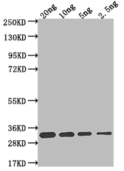
To create the L1R antibody, a rabbit is immunized with recombinant vaccinia virus L1R (2-183aa), inducing the rabbit's B lymphocytes to produce specific IgG antibodies. The polyclonal L1R antibody is then meticulously purified from the rabbit's serum using protein G techniques. The functionality of this L1R antibody has been validated in both ELISA and WB tests. This L1R antibody can react with the vaccinia virus L1R protein.

L1R, a myristylated late gene product of the vaccinia virus, plays an essential role in the formation of infectious intracellular mature virions (IMV). The L1R gene-encoding protein L1 is located on the surface of IMVs and beneath the envelope of the extracellular enveloped virus (EEV). L1 is also involved in infection, as it has been observed that antibodies targeting L1 can effectively hinder the virus's invasion of host cells in its IMV form.

Customer Reviews and Q&A

 Customer Reviews

There are currently no reviews for this product.

Submit a Review here

Target Background

Function
Envelope protein which probably plays a role in virus entry into the host cell. Is probably involved in the virus attachment to the host cell surface and associates with the entry/fusion complex (EFC). Needed for fusion and penetration of the virus core into host cell.
Subcellular Location
Virion membrane; Single-pass membrane protein.
Protein Families
Chordopoxvirinae L1 family
icon of phone
Call us
301-363-4651 (Available 9 a.m. to 5 p.m. CST from Monday to Friday)
icon of address
Address
7505 Fannin St., Ste 610, Room 7 (CUBIO Innovation Center), Houston, TX 77054, USA
icon of social media
Join us with

Subscribe newsletter

Leave a message

* To protect against spam, please pass the CAPTCHA test below.
CAPTCHA verification
© 2007-2024 CUSABIO TECHNOLOGY LLC All rights reserved. 鄂ICP备15011166号-1