Call us
301-363-4651 (Available 9 a.m. to 5 p.m. CST from Monday to Friday)
Recombinant antibodies are a type of monoclonal antibodies, which are generated in vitro from a synthetic gene without immunizing animals or cultivating hybridomas. After cloning the antibody genes into an expression vector, then transfected into an appropriate host cell line for antibody expression. Compared to traditional antibodies, recombinant antibodies result in superior lot-to-lot consistency, continuous supply, and animal-free advantages. As such, recombinant antibodies are being used more and more widely for scientific research, especially as a solution to addressing the repeatability crisis.
CUSABIO has been working on the development and production of recombinant antibodies since from 2020. Currently, CUSABIO has more than 1000 recombinant antibodies in stock, and most of CUSABIO recombinant antibodies had been validated in multipe applications. In order to better help our customers on research, CUSABIO also had successfully produced the recombiant antibodies specific for many difficult targets, such as: CLDN18.2, CLDN6, CCR8, SSTR2, DLL3, LY6G6D, ROR1, SEMA4D, GPC3, CD16A, etc.
● Advanced Phage Display Technology
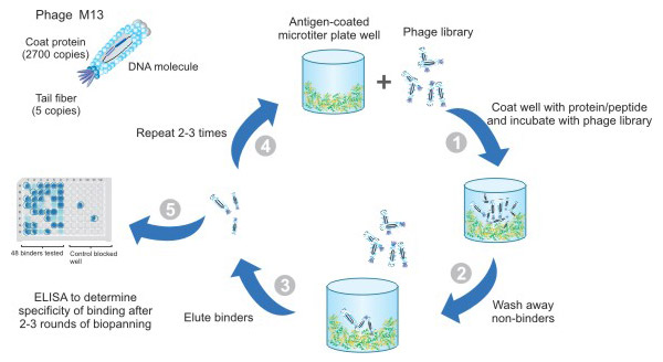
CUSABIO has been specialized in applying advanced phage display technologies for a wide range of service projects, especially development of recombinant antibody. Read more>>
● Various Types of Recombinant Antibody
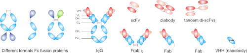
CUSABIO offers various types of recombinant antibody, including full-length antibodies, scFv, Fab, sdAb, isotype/subtype and various forms of Fc fusion proteins.
● Rich Application Types
CUSABIO's recombinant antibodies have been validated on multiple platforms, including WB, IF, IHC, FC, IP, ELISA, GICA and Neutralising.
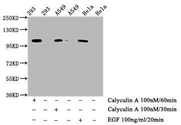
Phospho-ERN1 (S724) Antibody (CSB-RA007795A724phHU)
Positive WB detected in: 293 whole cell lysate, A549 whole cell lysate, Hela whole cell lysate (treated with Calyculin A or EGF)
All lanes: Phospho-ERN1 antibody at 0.75μg/ml
Secondary
Goat polyclonal to rabbit IgG at 1/50000 dilution
Predicted band size: 110 kDa
Observed band size: 110 kDa
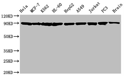
HSP90AA1/HSP90AB1 Antibody (CSB-RA010802A0HU)
Positive WB detected in: Hela whole cell lysate, MCF-7 whole cell lysate, K562 whole cell lysate, HL-60 whole cell lysate, HepG2 whole cell lysate, A549 whole cell lysate, Jurkat whole cell lysate, PC3 whole cell lysate, Rat brain tissue
All lanes: Hsp90 alpha + beta antibody at 1.25μg/ml
Secondary
Goat polyclonal to rabbit IgG at 1/50000 dilution
Predicted band size: 90 KDa
Observed band size: 90 KDa
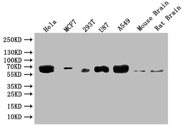
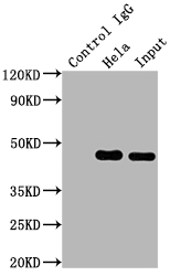
GBA Antibody (CSB-RA869334A0HU)
Positive WB detected in: Hela whole cell lysate, MCF7 whole cell lysate, 293T whole cell lysate, U87 whole cell lysate, A549 whole cell lysate, Mouse Brain tissue, Rat Brain tissue
All lanes: GBA antibody at 1:500
Secondary
Goat polyclonal to rabbit IgG at 1/50000 dilution
Predicted band size: 60 kDa
Observed band size: 60 kDa
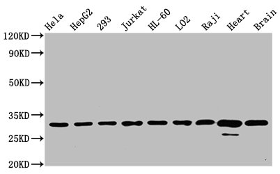
VDAC1 Antibody (CSB-RA025821A0HU)
Positive WB detected in: Hela whole cell lysate, HepG2 whole cell lysate, 293 whole cell lysate, Jurkat whole cell lysate, HL-60 whole cell lysate, LO2 whole cell lysate, Raji whole cell lysate, Rat heart tissue, Mouse brain tissue
All lanes: VDAC1 antibody at 0.7μg/ml
Secondary
Goat polyclonal to rabbit IgG at 1/50000 dilution
Predicted band size: 31 KDa
Observed band size: 31 KDa
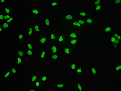
Histone H3.3 Antibody (CSB-RA010109A0HU)
Immunofluorescence staining of Hela cells with the antibody at 1:60, counter-stained with DAPI.
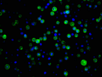
CD21 Antibody (CSB-RA005934A0HU)
Immunofluorescence staining of Raji cells with the antibody at 1:34, counter-stained with DAPI.
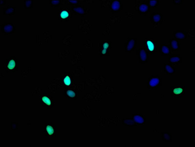
Histone H3.1 Antibody (CSB-RA010418A0HU)
Immunofluorescence staining of Hela cells with the antibody at 1:93, counter-stained with DAPI.
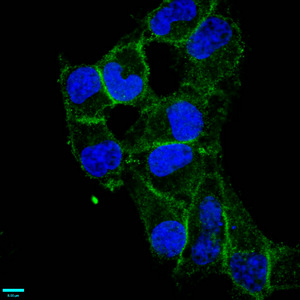
CD97 Antibody (CSB-RA004972A0HU)
Immunofluorescence staining of Hela cells with the antibody at 1:87.5, counter-stained with DAPI.
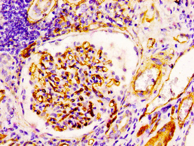
CD34 Antibody (CSB-RA004926A0HU)
IHC image of the antibody diluted at 1:100 and staining in paraffin-embedded human kidney tissue performed on a Leica BondTM system.
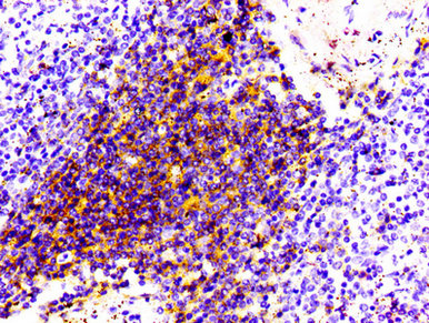
CD4 Antibody (CSB-RA004935A0HU)
IHC image of the antibody diluted at 1:100 and staining in paraffin-embedded human spleen tissue performed on a Leica BondTM system.
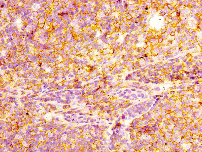
CD44 Antibody (CSB-RA004938A0HU)
IHC image of the antibody diluted at 1:100 and staining in paraffin-embedded human tonsil tissue performed on a Leica BondTM system.
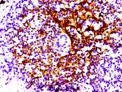
CD21 Antibody (CSB-RA005934A0HU)
IHC image of the antibody diluted at 1:100 and staining in paraffin-embedded human lung cancer performed on a Leica BondTM system.
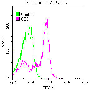
CD81 Antibody (CSB-RA004960A0HU)
Overlay histogram showing Jurkat cells stained with the antibody (red line) at 1:50. Control antibody (green line) was used under the same conditions. Acquisition of >10,000 events was performed.
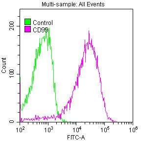
CD99 Antibody (CSB-RA004973A0HU)
Overlay histogram showing Jurkat cells stained with the antibody (red line) at 1:50. Control antibody (green line) was used under the same conditions. Acquisition of >10,000 events was performed.
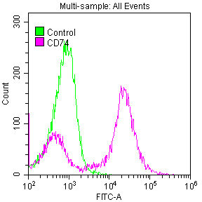
CD74 Antibody (CSB-RA004956A0HU)
Overlay histogram showing Raji cells stained with the antibody (red line) at 1:50. Control antibody (green line) was used under the same conditions. Acquisition of >10,000 events was performed.
CD163 Antibody (CSB-RA801238A0HU)
Overlay histogram showing Raw264.7 cells stained with the antibody (red line) at 1:50. Control antibody (green line) was used under the same conditions. Acquisition of >10,000 events was performed.
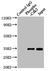
Phospho-CDK2 (Y15) Antibody (CSB-RA005061A15phHU)
Positive WB detected in:Hela whole cell lysate, 293 whole cell lysate(treated with Pervanadate or not)
All lanes:Phospho-CDK2 antibody at 0.8μg/ml
Secondary
Goat polyclonal to rabbit IgG at 1/50000 dilution
Predicted band size: 34 KDa
Observed band size: 34 KDa
MAP2K1 Antibody (CSB-RA957619A0HU)
Positive WB detected in: Hela whole cell lysate, 293 whole cell lysate, MCF-7 whole cell lysate, 293T whole cell lysate, A549 whole cell lysate, U251 whole cell lysate, Rat brain tissue
All lanes: MAP2K1 antibody at 1:2000
Secondary
Goat polyclonal to rabbit IgG at 1/50000 dilution
Predicted band size: 44, 41 kDa
Observed band size: 44 kDa
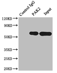
PAK2 Antibody (CSB-RA592787A0HU)
Positive WB detected in: 293 whole cell lysate, Jurkat whole cell lysate, Raji whole cell lysate, Mouse brain tissue, Rat brain tissue
All lanes: PAK2 antibody at 1:2000
Secondary
Goat polyclonal to rabbit IgG at 1/50000 dilution
Predicted band size: 59 kDa
Observed band size: 59 kDa
PKM Antibody (CSB-RA632595A0HU)
Positive WB detected in: SH-SY5Y whole cell lysate, Jurkat whole cell lysate, MCF-7 whole cell lysate, Hela whole cell lysate, Raji whole cell lysate, 293 whole cell lysate, Mouse brain tissue
All lanes: PKM antibody at 1:2000
Secondary
Goat polyclonal to rabbit IgG at 1/50000 dilution
Predicted band size: 58, 59, 57 kDa
Observed band size: 58 kDa
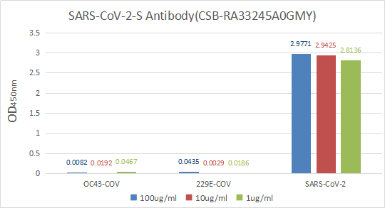
Immobilize various types of SARS proteins at concentration of 2μg/ml on solid substrate, then react with SARS-CoV-2-S Antibody at concentration of 100μg/ml, 10μg/ml and 1μg/ml. It shows the SARS-CoV-2-S Antibody (CSB-RA33245A0GMY) is specific for SARS-CoV-2-S1-RBD protein, without any cross-reactivity with HCoV-OC43, HCoV-229E.
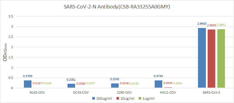
Immobilize various types of SARS proteins at concentration of 2μg/ml on solid substrate, then react with SARS-CoV-2-N Antibody at concentration of 100μg/ml, 10μg/ml and 1μg/ml. It shows the SARS-CoV-2-N Antibody (CSB-RA33255A0GMY) is specific for SARS-CoV-2-N protein, without any cross-reactivity with NL63-CoV, HCoV-OC43, HCoV-229E or HCoV-HKU1.
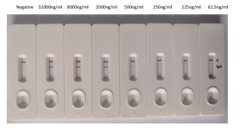
In the Colloidal Gold Immunochromatography Assay detection system, the background of antibody (CSB-RA33255A1GMY) is clean, the detection limit can be as low as 125ng/mL (8.75ng/0.07mL), and the sensitivity is very good.
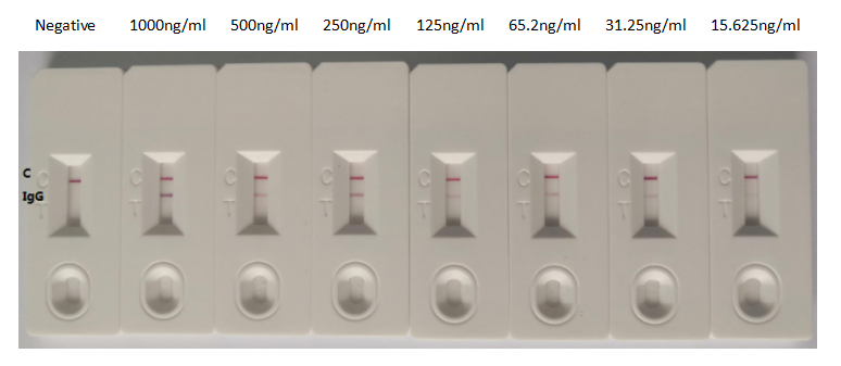
In the Colloidal Gold Immunochromatography Assay detection system, the background of antibody (CSB-RA33255A2GMY) is clean, the detection limit can be as low as 15.625ng/ml (1.09ng/0.07ml), and the sensitivity is very good
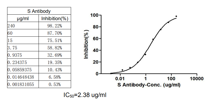
Binding signal of SARS-CoV-2-S1-RBD (CSB-YP3324GMY1) and ACE2 protein-HRP conjugate (CSB-MP866317HU) was inhibited by S Antibody (CSB-RA33245A1GMY) with the IC50 is 2.38 μg/Ml.
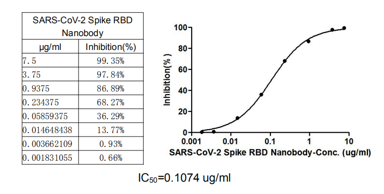
SARS-CoV-2 Spike RBD Nanobody (CSB-RA33245A2GMY)
This nanobody competitively prevented SARS-CoV-2-S1-RBD (CSB-YP3324GMY1) from binding to ACE2-HRP conjugate (CSB-MP866317HU). The inhibition efficacy of the SARS-CoV-2-S1-RBD/ACE2 binding was positively proportionally to the nanobody concentrations. It showed that the nanobody effectively inhibited the SARS-CoV-2-S1-RBD/ACE2 binding. And the IC50 of the nanobody is 0.1074 μg/mL.
● High Consistency from Batch To Batch
Ensuring reliable results with each experiment, our rigorous quality control processes maintain uniformity across all product batches.
● Continuity of Supply
With a robust supply chain, CUSABIO guarantees a steady and uninterrupted availability of products, supporting your long-term research needs.
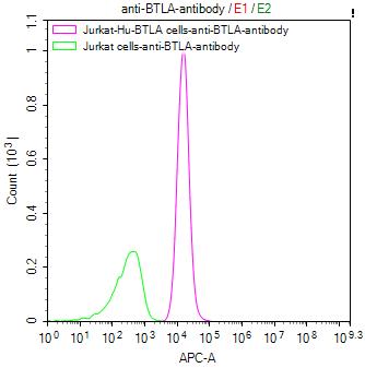
BTLA Recombinant Monoclonal Antibody
CSB-RA773799MA1HU
Untransfected HEK293T cells (green line) and transfected Human ENPP3 HEK293T Stable cells (red line) were stained with anti-ENPP3 antibody (2µg/1*106).
CD247 Recombinant Monoclonal Antibody
CSB-RA244537A0HU
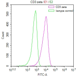
Overlay Peak curve showing Jurkat cells surface stained with CSB-RA244537A0HU (red line) at 1:50.
CD274 Recombinant Monoclonal Antibody
CSB-RA878942MA1HU
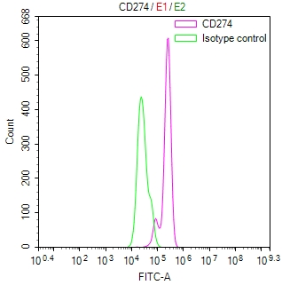
Overlay Peak curve showing Hela cells surface stained with CSB-RA878942MA1HU (red line) at 1:100.
CD40 Recombinant Monoclonal Antibody
CSB-RA004936MA1HU
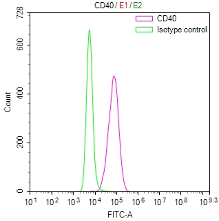
Overlay Peak curve showing THP-1 cells surface stained with CSB-RA004936MA1HU (red line) at 1:100.
CCR4 Recombinant Monoclonal Antibody
CSB-RA004843MA01HU
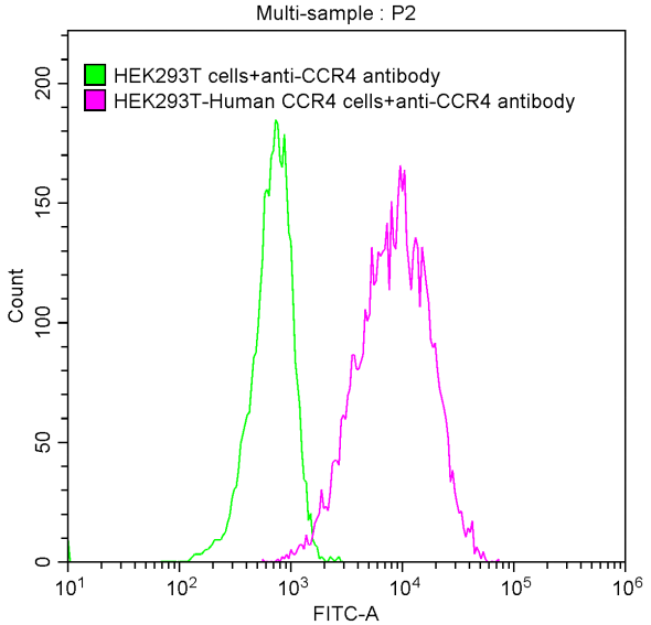
Untransfected HEK293 cells surface (green line) and transfected Human CCR4 HEK293 stable cells surface (red line) were stained with anti-CCR4 antibody(2µl/1*106 cells)
Claudin-18.2 Recombinant Monoclonal Antibody
CSB-RA005498A1HU
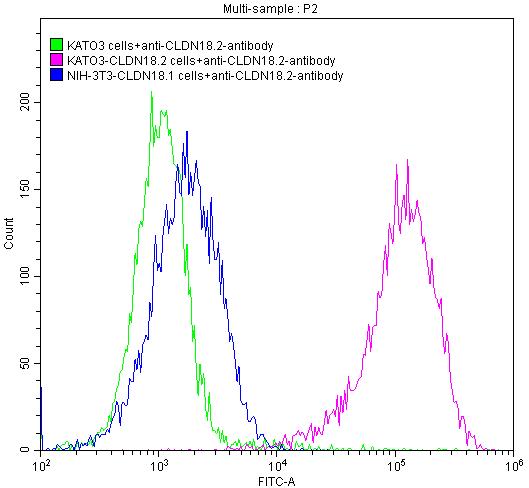
Untransfected KATO3 cells surface (green line), transfected Human CLDN18.2 KATO3 stable cells surface (red line) and transfected.
CD3E Recombinant Monoclonal Antibody
CSB-RA004931MA1HU
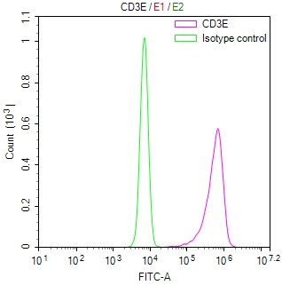
Overlay Peak curve showing JK cells surface stained with CSB-RA004931MA1HU (red line) at 1:100.
CD44 Recombinant Monoclonal Antibody
CSB-RA004938MA1HU
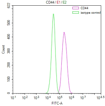
Overlay Peak curve showing Hela cells surface stained with CSB-RA004938MA1HU (red line) at 1:100.
S Antibody referenced in "A magneto-optical biochip for rapid assay based on the Cotton–Mouton effect of γ-Fe2O3@ Au core/shell nanoparticles", Journal of nanobiotechnology, 2021.
SARS-CoV-2 Spike RBD Nanobody referenced in "Potent antiviral activity of Agrimonia pilosa, Galla rhois, and their components against SARS-CoV-2", Bioorganic & Medicinal Chemistry, 2021.
HIF1A Antibody, CASP3 Antibody referenced in "PESV represses non-small cell lung cancer cell malignancy through circ_0016760 under hypoxia", Cancer cell international, 2021.
Phospho-ERN1 (S724) Antibody referenced in "The ER stress sensor inositol-requiring enzyme 1α in Kupffer cells promotes hepatic ischemia-reperfusion injury", Journal of Biological Chemistry, 2021.
CDK4 Antibody and CDK6 Antibody referenced in "Histone Deacetylase Inhibitor-Induced CDKN2B and CDKN2D Contribute to G2/M Cell Cycle Arrest Incurred by Oxidative Stress in Hepatocellular Carcinoma Cells Via Forkhead Box M1 Suppression", Journal of Cancer, 2021.
BIRC5 Antibody referenced in "LTBP4 Inhibits the Proliferation and Metastasis in Melanoma by Activating Hippo-YAP Signaling", Research Square, 2020.
A stable and highly specific antibody plays an important role in obtaining real and repeatable experimental results. It is a big headache if the antibody used in the experiment does not work. And what's even worse is that you use the antibodies to repeat the same experiment but get inconsistent results.
It is estimated that the annual loss in research funding due to the use of antibodies without enough validation is about $ 800 million worldwide. At the same time, it has caused countless experimental failures, and a waste of precious samples and researchers'youth.
Nowadays, the antibody validations information provided by antibody manufacturers and academic articles is very helpful for you to select high-quality antibodies. However, there is still an inevitable problem: whether it is polyclonal antibodies extracted from animal serum or monoclonal antibodies produced by hybridomas, there are more or fewer differences between batches. And recombinant antibodies may finally solve this problem.
Recombinant antibodies refers to the application of molecular cloning and gene mutation technology to transform a gene coding sequence of an antibody to produce superior performance of antibodies, also known as genetically engineered antibodies. Comparing with monoclonal antibodies generated in vivo of living animals through hybridoma technology, recombinant antibodies are a type of monoclonal antibodies which are produced in vitro by using synthetic genes.
In recent years, the use of recombinant antibodies is becoming increasingly wider and deeper in both therapeutics and diagnostics. With significant advantages over conventional antibodies, recombinant antibodies become more and more favored. The most important point is that recombinant antibodies can maximize the humanization to solve the heterogeneity between different species.
Generally speaking, recombinant antibodies can be divided into three classes, including chimeric antibody, humanized antibody, fully humanized antibody and small molecular antibody.
Compared with traditional antibodies, recombinant antibodies have the advantages of being irresistible, which is one of the reasons for their gradually emerging. Here, we present the advantages one by one as follows:
Because recombinant antibodies have these unprecedented advantages, recombinant antibodies fulfill a large spectrum of functions spanning from research to diagnosis and treatment therapies for various diseases. Their specificity and low immunogenicity make them a great alternative to traditional forms of treatment, increasing the accuracy of targeting specific molecules and avoiding adverse side effects.
Additionally, for recombinant antibodies production, it follows principally similar workflow. Generally, it consists of five steps: the construction of antibody gene library, phage display, the separation of antibodies, the modification of the isolated antibodies, and the scaling up of the production of selected antibodies in the cell culture expression system.
Although the processes of recombinant antibodies production are same as workflow, we need to recognize that there are several obstacles to the production of recombinant antibodies, such as lower antibody production, and highly trained and experienced technicians. Due to the complexity and intensive high technology of recombinant antibody production, most scientists need to obtain them from outsourcing companies.
Read the article, The Overview of Recombinant Antibody, for more information.