Call us
301-363-4651 (Available 9 a.m. to 5 p.m. CST from Monday to Friday)
PMEL, or Premelanosome Protein, is a protein expressed in melanocytes and encoded on chromosome 12 mainly in humans.PMEL plays a key role in the formation and function of melanosomes and is involved in the formation of fibrillar structures within melanosomes that contribute to melanin synthesis and distribution.PMEL proteins achieve their functions through their different structural domains, including signal peptides, transmembrane domains, and cytoplasmic domains, as well as undergoing post-translational modifications such as protease cleavage and glycosylation. In a biological sense, PMEL is essential for the maintenance of skin and eye pigmentation, and it may also play a role in the development of certain skin diseases such as melanoma, making it an important target for the study of skin pigmentation and related diseases.
● Recombinant Human Melanocyte protein PMEL (PMEL), partial
Validated Data
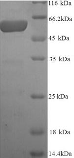
(Tris-Glycine gel) Discontinuous SDS-PAGE (reduced) with 5% enrichment gel and 15% separation gel.
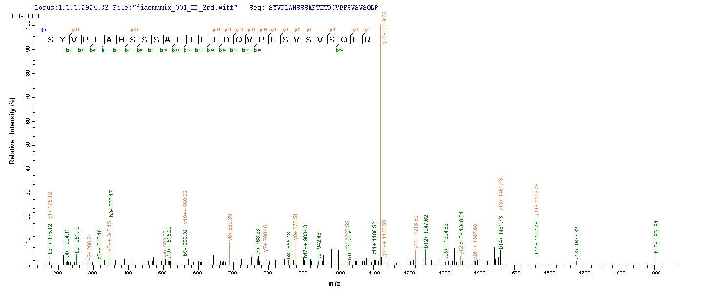
Based on the SEQUEST from database of Yeast host and target protein, the LC-MS/MS Analysis result of CSB-YP021324HU could indicate that this peptide derived from Yeast-expressed Homo sapiens (Human) PMEL.
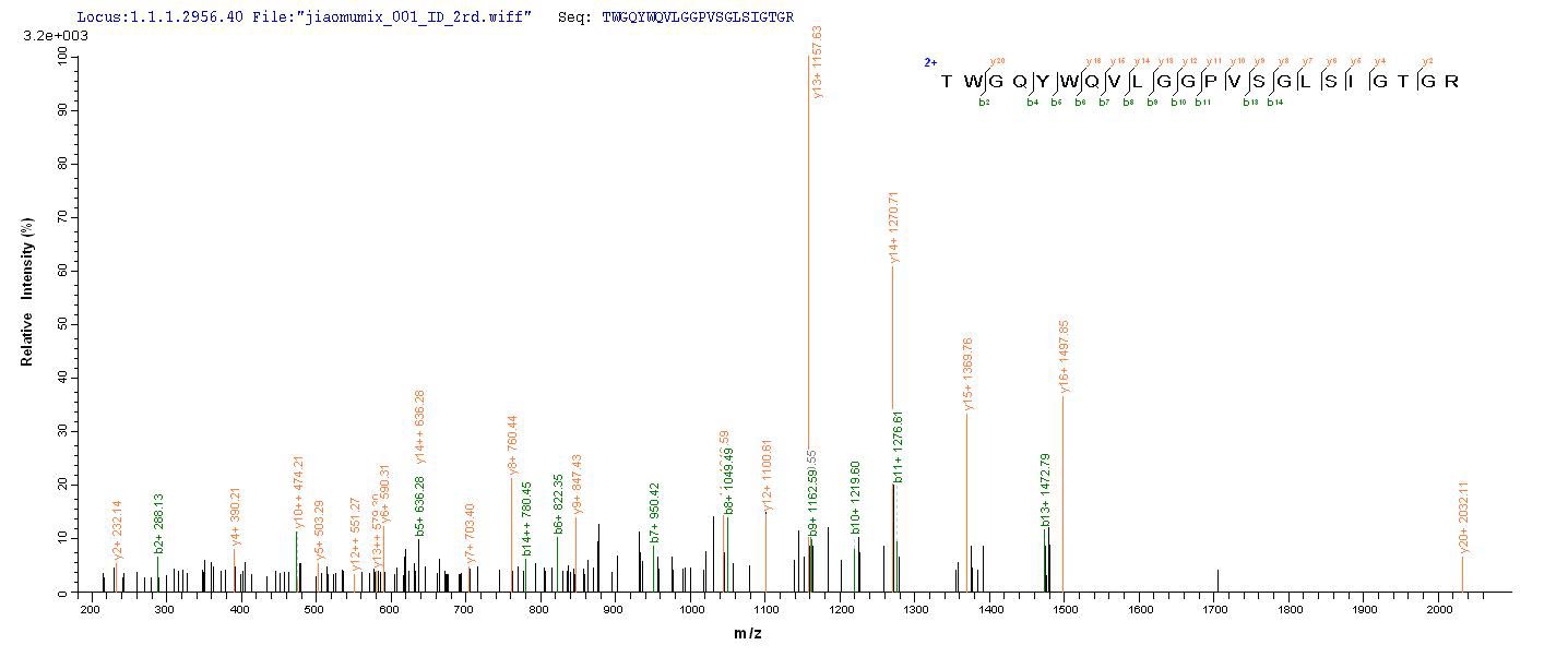
Based on the SEQUEST from database of Yeast host and target protein, the LC-MS/MS Analysis result of CSB-YP021324HU could indicate that this peptide derived from Yeast-expressed Homo sapiens (Human) PMEL.
Validated Data
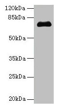
Western blot
All lanes: PMEL antibody at 14µg/ml + Jurkat whole cell lysate
Secondary
Goat polyclonal to rabbit IgG at 1/10000 dilution
Predicted band size: 71, 61, 67 kDa
Observed band size: 71 kDa
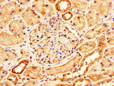
IHC image of CSB-PA021324LA01HU diluted at 1:200 and staining in paraffin-embedded human kidney tissue performed on a Leica BondTM system. After dewaxing and hydration, antigen retrieval was mediated by high pressure in a citrate buffer (pH 6.0). Section was blocked with 10% normal goat serum 30min at RT. Then primary antibody (1% BSA) was incubated at 4°C overnight. The primary is detected by a biotinylated secondary antibody and visualized using an HRP conjugated SP system.
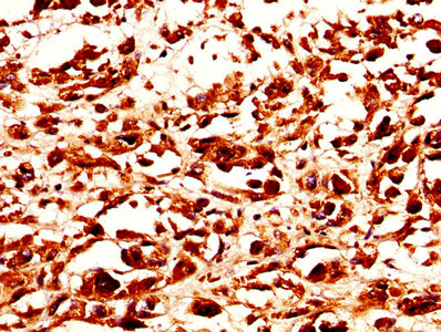
IHC image of CSB-PA021324LA01HU diluted at 1:200 and staining in paraffin-embedded human melanoma performed on a Leica BondTM system. After dewaxing and hydration, antigen retrieval was mediated by high pressure in a citrate buffer (pH 6.0). Section was blocked with 10% normal goat serum 30min at RT. Then primary antibody (1% BSA) was incubated at 4°C overnight. The primary is detected by a biotinylated secondary antibody and visualized using an HRP conjugated SP system.
The following PMEL reagents supplied by CUSABIO are manufactured under a strict quality control system. Multiple applications have been validated and solid technical support is offered.
PMEL Antibodies for Homo sapiens (Human)
| Code | Product Name | Species Reactivity | Application |
|---|---|---|---|
| CSB-PA021324LA01HU | PMEL Antibody |
Human | ELISA, WB, IHC |
| CSB-PA021324LC01HU | PMEL Antibody, FITC conjugated |
Human | |
| CSB-PA021324LD01HU | PMEL Antibody, Biotin conjugated |
Human | ELISA |
PMEL Proteins for Homo sapiens (Human)
| Code | Product Name | Source |
|---|---|---|
| CSB-YP021324HU | Recombinant Human Melanocyte protein PMEL (PMEL), partial |
Yeast |
| CSB-EP021324HU | Recombinant Human Melanocyte protein PMEL (PMEL), partial |
E.coli |
| CSB-BP021324HU CSB-MP021324HU CSB-EP021324HU-B |
Recombinant Human Melanocyte protein PMEL (PMEL), partial, |
Baculovirus Mammalian cell In Vivo Biotinylation in E.coli |